Speakers
Description
Summary
Neutze et al. (1) first suggested the use of XFELs for biomolecular imaging since the timescale of X-ray pulses irradiating membrane-protein samples (~ 5-50 femtoseconds) is such that nuclear damage may be avoided. Further consideration has also been paid to damage to the electronic structure of such biomolecules under intense radiation (2).
Coherent Diffractive Imaging (CDI) is an imaging technique that has been developed to image objects that are non-crystalline (3)(4) with X-rays. The object structure is reconstructed from the diffraction data image using phase-retrieval algorithms (5)(6). It has been demonstrated that appropriate real-space constraints (information about the sample object such as its size) or implementation of knowledge about the wavefield in phase-retrieval algorithms improves the reconstruction of the sample (7)(8).
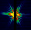
The collection of diffraction data and analysis from single molecules and small two-dimensional membrane protein crystals using XFEL pulses has been considered (9)(10)(11). We have performed diffraction simulations for submicron membrane protein crystals incorporating the X-ray intensities supplied at LCLS, Stanford. We seek to develop implement CDI techniques for resolving high resolution information from this data for experiments to be performed in the near future.
(1)Neutze et al., Nature, 2000
(2)Hau-Riege et al.,Phys Rev B., 2004
(3)Sayre, Acta Crystallographica, 1952
(4)Miao, Nature, 1999
(5)Gerchberg, Saxton, Optik, 1972
(6)Feinup, Applied Optics, 1978
(7)Nugent et al., Phys Rev Lett., 2003
(8)Quiney et al., Nature Physics, 2008
(9)Ourmazd et al., Nature Physics, 2009
(10)Spence, et al., Phys Rev Lett,2004
(11)Mancuso et al., Phys Rev Lett., 2009

